Decoding the developing brain in Seoul: Dr Hosna Asma-Ull’s autism research
What can a brain scan reveal about a child’s development? Can artificial intelligence help researchers identify early differences and give doctors more time to respond?
Decoding the developing brain in Seoul: Dr Hosna Asma-Ull’s autism research
What can a brain scan reveal about a child’s development? Can artificial intelligence help researchers identify early differences and give doctors more time to respond?
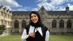
Dr Hosna Asma-Ull, a Bangladeshi researcher based in South Korea, is exploring these questions as a postdoctoral researcher in the Department of Radiology at Asan Medical Center in Seoul.
She uses artificial intelligence (AI) and deep learning to analyse children’s MRI scans and study brain development, including patterns relevant to autism research.
Hosna completed her SSC at Khilgaon Girls High School and HSC at Motijheel Ideal College before moving to South Korea to study at Sookmyung Women’s University. After her BSc, she returned to Bangladesh to teach computer science at Stamford University Bangladesh. She later returned to South Korea for her MSc and PhD before beginning her current research.
Her work explores whether measurable differences in the developing brain can help researchers better understand developmental outcomes. But she cautions against confusing detection with diagnosis.
“Detecting something is not the same as being able to do anything about it,” she said.
Understanding a changing brain
A child’s brain develops rapidly during infancy, making it difficult to establish a single standard for every scan.
“Every organ changes over a lifetime, but few change as fast as a child’s brain,” Hosna said. “Its structure shifts month by month in the early years, which means ‘normal’ is not a fixed reference but a moving one. What is typical at six months is not typical at eighteen.”
Using deep learning models developed largely with Python, she analyses MRI scans to identify brain structures and measure their characteristics.
One area of her research is brain-age estimation, which compares a scan with patterns typically seen at different ages.
“A brain age works the same way, except the measuring tape is an MRI scan,” she said.
A difference between estimated and actual brain age may warrant further investigation, but it cannot independently diagnose autism.
“Autism is not one condition with one signature,” Hosna said. “Children develop along enormously varied paths, and a measurement that means one thing in one child may mean nothing in another.”
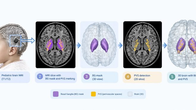
She also studies perivascular spaces, tiny fluid-filled spaces surrounding blood vessels in the brain. In a study published in BMC Medical Imaging, Hosna and her colleagues examined whether these spaces in neonatal brain scans were associated with neurodevelopmental outcomes at 24 months.
For her, AI remains a research tool, not a replacement for medical judgement.
“We are still a long way from a cure for autism, and in truth ‘cure’ may not be the right word at all,” she said.
“What early detection offers is time,” she added.
Teaching AI with synthetic brain scans
Medical AI research often faces a shortage of large, accurately labelled datasets, particularly for rare conditions. Labelling scans requires specialist expertise, while privacy requirements can limit access to medical records.
“The data exists, the expertise exists, and yet the datasets we can actually assemble are small,” Hosna said.
Her research explores whether generative AI can help by creating synthetic MRI images that do not belong to real patients. Using diffusion models, researchers can generate artificial fetal and neonatal brain scans showing conditions such as ventriculomegaly, an enlargement of fluid-filled spaces in the brain, and microcephaly, where a child’s head is smaller than expected.
These images could provide additional examples of rare conditions, but their value depends on whether they improve AI performance on real patient scans.
“If adding synthetic pathological cases makes a segmentation or classification model perform better on real patient scans, particularly for conditions that are too rare to learn from otherwise, then the images have earned their place,” she said. “If not, they are only pictures.”
Bringing the research closer to Bangladesh
Hosna believes medical AI must account for differences between populations, scanners and imaging methods. A model trained mainly on Korean, European or North American children may not perform equally well on Bangladeshi children.
“A model is only as general as the children it learned from,” she said.
She argues that Bangladeshi researchers need to develop and test local datasets rather than assume models developed elsewhere will work equally well here.
Access to scientific literature is another challenge. Hosna has proposed that the University Grants Commission negotiate national access to major scientific publishers, allowing students and researchers to build on existing studies.
“You cannot build datasets without knowing what a good dataset looks like. You cannot design studies without knowing what has already been tried. Before anything else, we have to let people see the horizon,” she said.
She also wants to explore AI tools that could improve low-quality MRI, CT and X-ray images produced by older or less expensive equipment in Bangladeshi hospitals. Such tools could help doctors work with existing resources, but they would need rigorous testing on local images and could not replace radiologists.
“Most medical artificial intelligence is built on the assumption of excellent data, which quietly means it is built for wealthy hospitals,” she said. “This would work the other way around.”
Her current five-year, Korean government-funded project uses datasets from Korea, Switzerland and the United States to investigate why methods that work on one dataset may fail on another. The aim is to develop more reliable approaches that work across populations and with modest computing resources.
Learning to ask better questions
Hosna’s path into medical imaging was not direct. During her MSc and PhD, she worked with images of nearly 10 organs and tissues, including blood vessels, the retina, knee cartilage and skin lesions. This taught her that methods suited to one type of tissue may fail when applied to another.
“A blood vessel does not fail the way cartilage fails. A retina does not photograph the way skin does,” she said.
She estimates that she read around 300 papers during her doctoral years, often sketching ideas on their blank backs to work through difficult concepts. For her, research involves understanding what has already been tried, identifying why a method fails and finding a better approach.
The answers to questions about autism and early brain development remain uncertain. AI cannot yet explain why a child develops differently or predict exactly what that child will need.
By studying how the brain changes and developing methods that account for differences between populations, Hosna hopes to help researchers better understand early development.
The goal is not simply to detect a difference in a scan, but to establish whether that information can help doctors make better decisions and give children and their families more time to respond.